Introduction
Alkaptonuria is a rare metabolic disorder of autosomal recessive inheritance, resulting from deficiency of homogentisic acid oxidase enzyme in the metabolism of phenylalanine and tyrosine.1, 2, 3, 4, 5, 6, 7, 8, 9, 10 The incidence is 1 in 25,000 to 1 in 1,000,000 individuals. 1, 4, 6, 7, 8, 9, 10 Homogentisic acid undergoes oxidation to form a brown black pigment, which results in the characteristic cola colored urine. 1 In infancy, complain of dark staining of nappies may be present.4 Ochronosis manifests when degenerative changes affect joint cartilage due to black pigment deposition in connective tissues following homogentisic acid polymerization. 1, 4, 10 The patients present usually with clinical symptoms only in the 3rd to 4th decade of life with stiffness and pain in low back as their initial symptom, when lumbosacral spine radiographs reveal decrease in the height of intervertebral discs, syndesmophyte, loss of lumbar lordosis mimicking AS clinically and radiologically. 1, 5, 9, 10 As the disease progresses in three to four years, from the initial presentation the typical radiological signs of calcification of intervertebral discs develops. 1, 4 These patients have propensity to undergo progressive osteoarthritic changes and multi-system involvement. Since treatment is palliative, it is imperative to diagnose the condition early, educate the patient to improve quality of life.
We report herein occurrence of the disease in two brothers with early to late onset ochronosis, resembling AS, and emphasize the importance of differentiating from AS and its early recognition.
Case Report
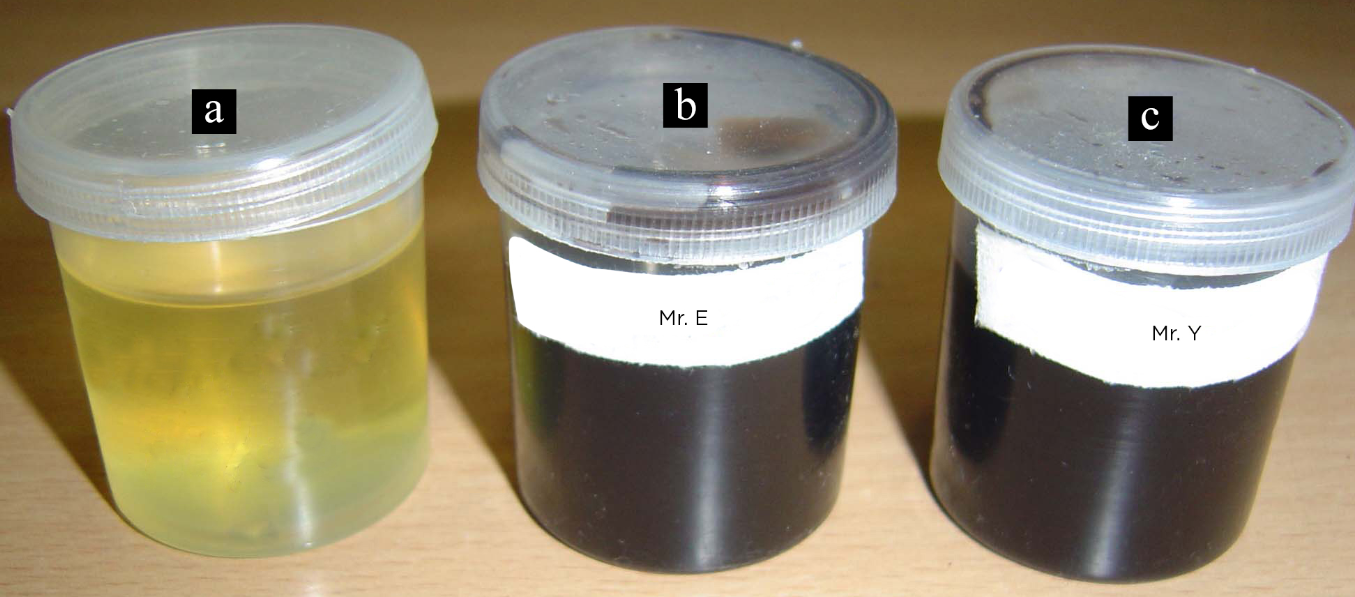
Elder sibling (Mr E), 33-years old male, of Dravidian origin, was suffering from complaints of low back pain and stiffness for the past four years. There was no history of night pain, although morning stiffness affects him for a short duration of less than thirty minutes. There was no history of trauma, radicular pain and associated systemic illness. His clinical examination revealed mild dorsal kyphosis, restricted chest expansion and reduced lumbar spine flexion confirmed by Schoeber's test (Figure 1a). The face had darker complexion when compared to the rest of the body. He had black scleral spots in between the cornea and outer canthus in both the eyes, which was confirmed to be only pigmentation in scleral substance (Figure 1b and 1c). His lumbosacral spine radiographs done four years earlier at the age of 29-years revealed reduced inter-vertebral discs spaces, osteophytosis and decreased lumbar lordosis (Figure 2a and 2b). A provisional diagnosis of AS was considered and management initiated by local physician with no relief. However, the repeat radiographs of the lumbosacral spine at the time of his presentation revealed calcification of the inter-vertebral discs, grossly reduced inter-vertebral disc spaces and loss of lumbar lordosis (Figure 2c and 2d). Radiographs of pelvis with both hips did not reveal any significant abnormality in the sacroiliac joints or the hip joints. His ESR was normal and HLA B-27 was negative. There was history of blackish brown staining of diapers and toilet pots since childhood. Urine turned blackish brown on exposure to air in comparison to a normal urine sample (Figure 3) and additional urinary tests with benedicts solution, ferric chloride and sodium hydroxide test were positive. Homogentisic acid quantification in urine was high. Magnetic resonance imaging (MRI) of the spine shows decreased height of the intervertebral discs and calcifications of the discs with degenerative changes in L1-2, L2-3, L3-4, and L4-5 levels (Figure 4a, 4b and 4c). Echocardiogram did not reveal any significant abnormality in the valves and wall motion. Global ejection fraction was 53%. He was put on high doses of vitamin C, educated about his possible outcomes and asked to maintain a strict regular physiotherapy and spine exercises regime. An inquiry into his family background had no evidence to suggest consanguineous marriage though his only other younger brother has complaints of low back pain.
Younger sibling (Mr. Y), a 30-year old male worked as an engineer and complaints of mild pain and stiffness of lower back from four months. His physical examination demonstrated decreased lumbar lordosis with stiffness of the lumbosacral spine with pain on flexion and extension. Findings on neurological examination were normal. Lumbo-sacral spine radiographs revealed decreased intervertebral disc spaces and syndesmophytes (Figure 5a and 5b). His ESR was normal, HLA B-27 was negative, urine turned black on standing (shown in Figure 3) and MRI showed degenerative changes at multiple lumbar disc level L2-3, L3-4, L4-5 and L5-S1 levels. The same treatment protocol was followed as for his elder sibling.
Figure 1
Clinical photograph ofMr. E with limited spinal flexion with white arrow showing span of expansion on, 1a and black scleral pigmentation of right and left eye in, 1b and 1c respectively.

Figure 2
Radiograph of lumbar spine ofMr. E, lateral and anteroposterior view in, 2a and 2b with white arrow marked at early osteophytes at L1-L2, L2-L3, L3-L4, L4-L5 intervertebral disc spaces. Radiograph of lumbar spine at a four-year interval lateral and anteroposterior view in 2c and 2d with black arrows showing syndesmophytes, marginal bridging and wafer like disc calcifications.

Figure 3
Photograph of containers with urine of normal colour marked‘a’ and black discoloured urine on exposure to air in container ‘b’ from Mr. E and container ‘c’ from Mr. Y.

Discussion
Alkaptonuria is the first disorder in humans to be found to confirm to the principles of mendelian autosomal recessive inheritance. Homogentisic acid oxidase, is an important enzyme involved in the metabolism of the aromatic amino acid phenylalanine and tyrosine. It turns black on oxidation and the metabolic products gets accumulated in various tissues like cheeks, axilla, genital regions, sclera, skin, ear, larynx, nail beds, joint cartilages, inter-vertebral disc, and tendon leading to ochronosis. 1, 2, 4, 5, 7, 8, 9 The urine tends to turn dark on prolonged exposure to air and there is history of dark staining of nappies in childhood. 1, 4, 6, 7, 8, 9 The reducing properties of urine when an alkali was added to an affected patient urine sample caused darkening of the urine. 4
Higher frequency is seen in Slovakia, Santo Domingo and certain middle east countries, where incidence approaches 19-25 per 500,000 inhabitants and elsewhere 1 per 10 million. 6 Reports of occurrence among siblings or among parents and children are infrequent. 6, 7, 8 On an average 1 in 4 siblings are affected, the risk being greater with consanguineous parents. In communities with numerous consanguineous marriages the incidence is higher. Men are commonly affected more than women, in ratio of about 2:1. 2
The symptoms of ochronotic spondyloarthropathy usually begin in late 30's with low back pain and stiffness. 2, 6 The two brothers present in their early 30’s with similar complaints. In the early phase of alkaptonuric spondylosis low back pain with sciatica may be the early symptom with loss of lumbar lordosis, and thoracic kyphosis becomes prominent. Clinically patients with ochronosis may present with less severe back pain than those with AS. Sacroiliac joints are rarely involved in ochronosis. The cervical spine is rarely involved although there may be forward protrusion of the head as a result of neck deformity, movements of the cervical spine usually remain free. Chest expansion may be decreased because of the involvement of costal and costovertebral joints.
There are reports on acute nucleus pulposus rupture of disc, fracture of osteoporotic ochronotic spine, cauda equina syndrome and acquired spinal canal stenosis in alkaptonuric ochronotic spondyloarthropathy. 10 Laminectomy, vertebroplasty and kyphoplasty are often required.10
Ochronosis affects many body systems. Dermatological and ophthalmological involvement appears in approximately 70% of the patients.2, 5 Some patients may develop aortic stenosis or mitral valve involvement due to pigment deposition.2 Renal and prostatic calculi have also been reported commonly.1, 2 Tendon ruptures as in Achilles tendon and patellar tendon have been reported.2, 7, 10 Ochronotic arthritis is characterized by progressive degenerative arthropathy mainly affecting axial weight bearing joints associated with extra articular manifestations. Involvement of large joints usually peripheral such as knees, hips, shoulder may occur with pain and limitation of motion being the common clinical features of the peripheral ochronotic arthropathy.2 Analyses have showed that procedures like joint replacement, cardiac valve replacement, renal stone surgery may be required for the management.7
High clinical suspicion is necessary when assessing low back pain. The assessment of ESR, serum alkaline phosphatase, blood sugar levels, C-reactive protein, haematocrit, white blood cell count, serum electrophoresis levels are required for further evaluation. Homogentisic acid excretion in the urine was higher, approximately three times than normal values. Urine color change upon alkalization is unpredictable and not specific for alkaptonuria. 10 Homogentisic acid (HGA) can be detected specifically by paper chromatography and quantitatively by the iodometric test. 9 Chromatographic techniques are necessary to reliably confirm the presence of HGA. Measurement of HGA in random urine is sufficient for establishing the diagnosis of alkaptonuria. 10 When alkalinized urine is placed on photographic paper, the paper turns dark.
Radiographic changes may precede the onset of symptoms. In the spine, the first changes are narrowing of disc spaces, commencing in the thoracolumbar region. The vertebral surfaces of the discs become increasing radio opaque due to wafer like calcification. Calcification of intervertebral discs of the spine is the pathognomic radiological sign of ochronosis. However, in contrast to AS syndesmophytes, annular ossification or bamboo spine pattern, does not occur. There is progressive disintegration and ossification of the nucleus pulposus. Contrasting with the radio opaque discs the vertebral bodies become osteoporotic. 1 Massive osteophyte formation develops at the vertebral margins and intervertebral bony bridging may occur. 1 A reactive spondylosis, halisteresis of the bones and pseudo block vertebrae are also frequent. These changes resemble with AS. Many of these changes may be interpreted as those of AS, but the striking density of the intervertebral discs combined with the usual almost complete absence of ligamentous ossification, central disc calcification, a maintained disc space, syndesmophytes and a ligamentous calcification differentiates the conditions. 1
On MRI the striking feature is the multiple levels of discs prolapse with the uniformly prominent low signal on T2 weighted images in all the discs, suggesting generalized degeneration. Nuclear imaging has little to offer in diagnosis.
Differential diagnosis includes degenerative disc disease, non-specific osteoarthritis, chondrocalcinosis, peri-articular calcification, osteo-chondromatosis.5 Differentiation of these diseases is based on history, clinical examination, laboratory findings, and radiologic appearance. 3
In degenerative disc disease vacuum phenomenon is related to cartilage brittleness as in peripheral joints. In osteoarthritis the spinal involvement is greatest in the lumbar and cervical regions. Disc calcification and osteophytic changes are unusual. 8 Progressive stiffening of the whole spine does not occur. Periarticular calcification may be a distinguishing feature in ochronotic arthritis. 3
In rheumatoid arthritis, the ESR is raised. Specific agglutination test may be positive. Cervical spine involved commonly with limitation of neck movement.3 Small joints usually spared in ochronotic arthropathy. 8
In AS, pain is more severe, cervical rigidity more marked and the ESR is usually high. AS is associated with thin and vertical syndesmophytes, severe apophyseal facet joint movement, erosion and fusion of SI joints. 3, 5
Discal calcification in the spine can also mimic hyperparathyroidism, haemochromatosis, amyloidosis, diffuse idiopathic skeletal hyperostosis (DISH), acromegaly, tuberculosis of spine, and in post traumatic spine conditions as well as in asymptomatic individuals. 1, 2, 5
There is no effective specific treatment. 3, 6, 7, 8, 9, 10 Treatment is usually conservative. 8 We followed a symptomatic approach, including treatment of pain, physiotherapy and education of the patient for a home exercise program to improve his quality of life. 3, 7 Awareness and early detection help in allaying anxiety. After fourth decade, monitoring and surveillance of cardiac and renal parameters is desirable. 4, 10
Vitamin-C, folic acid and cortisone have been tried without effect. 6 Vitamin C modifies the urinary excretion of HGA and decreases binding of ochronotic pigments to connective tissues. 6, 7 However, there is no definitive evidence for a retardation of cartilage damage with vitamin C in established ochronosis. 8, 10 Restriction of protein may improve symptoms however no significant change with a low protein diet is seen. 9, 10 Proposed treatment with nitisinone, a triketone herbicide has failed to show any promising results. The gene defect responsible for AKU was mapped to chromosome 3q13.33. 10 Genetic advances offer hope that corrective measures are forthcoming.
Alkaptonuria is usually compatible with a normal life span. Although, ochronotic arthropathy is considered to be a model of degenerative disease, it is often aggressive, with rapid destruction culminating in an end stage disease requiring joint prosthesis. Patient may be severely crippled and the benign nature of the condition has been mostly overemphasized. Alkaptonuria is a rare disorder presenting commonly with low back pain, which needs early recognition by clinicians and public alike. Lifestyle counselling of siblings with the same gender displaying differing severity of alkatonuric spondyloarthropathy is suggested for adopting activities and occupation that minimizes joint loading. 10 Continued education to emphasize the recognition of the dark urine, auricular and scleral pigmentation and radiological assessment as simple diagnostic clues will help in early recognition.


