- Visibility 362 Views
- Downloads 50 Downloads
- Permissions
- DOI 10.18231/j.ijor.2023.018
-
CrossMark

Isolated first metatarsal tubercular osteomyelitis of the foot in a boy: A case report with review of literature
- Author Details:
-
 Javed Ahmad
-
Mohit Kumar Singh
-
Brij Mohan Patel *
-
Vivek Kumar Shrivastava
-
Ajeet Kumar Yadav
Abstract
Isolated tubercular lesions of the metatarsal bones are rare, with an incidence of less than 0.5%. Because of ambiguous signs and symptoms, and reasonably normal laboratory investigations, these lesions pose a diagnostic challenge and there are chances of misinterpretation of radiographic images. We present a case of tubercular osteomyelitis of the first metatarsal of right foot in a 9-year-old boy. The diagnosis was initially made using plain X-ray, which was supported by magnetic resonance imaging and confirmed by the histopathological examination of the resected samples and microbiological culture combined with polymerase chain reaction. The patient was managed with open biopsy and curettage followed by a prolonged course of anti-tubercular treatment. The patient showed complete resolution of symptoms with no sign of recurrence.
Introduction
Tuberculosis has been known to humans since antiquity and continues to pose a serious health problem in developing and underdeveloped nations such as India.[1] Extrapulmonary tuberculosis affecting the bones and joints has come under the limelight again in recent times due to the changing epidemiology of the disease as a result of the increasing incidence of HIV/AIDS, multiple drug-resistant tuberculosis, and use of immunosuppressive drugs, among other factors. [2] Extrapulmonary involvement can occur alone or in conjunction with a pulmonary focus, as in the case of patients with disseminated tuberculosis, although the primary focus is always in the lungs.
Compared to the pulmonary form of tuberculosis, osteoarticular tuberculosis is rare, and difficult to diagnose, especially when it affects atypical areas.[3], [4] The skeletal system is affected in roughly 1% to 3% of all extrapulmonary tuberculosis cases. The spine and hip are the most often implicated locations in cases of skeletal tuberculosis. Only 10% of all cases of skeletal tuberculosis occur in the ankle and foot.[2], [3], [4], [5], [6] The calcaneum followed by the cuboid and talus are the most frequently involved sites. The involvement of an isolated metatarsal bone has rarely been reported in the literature.[4], [5], [6] Most of these reported cases have been at the extremes of age ranges.[7], [8], [9]
Because of ambiguous symptoms and lack of knowledge about the illness at atypical sites, the diagnosis of tuberculosis of the foot is frequently missed, which can delay the start of treatment. Pain and swelling in the affected bone or joint are the only initial symptoms. Later, widespread edema, discharging sinuses, and pathological fractures may occur. Plain radiographs are normal in the initial stages; however, computed tomography scan and magnetic resonance imaging may be useful for localizing the lesions.[6] Radiological images can mimic those observed in conditions, such as pyogenic osteomyelitis, Brodie's abscess, and neoplasms such as an aneurysmal bone cyst or a giant cell tumor, making it is necessary to confirm the diagnosis using Ziehl–Neelsen staining, culture sensitivity testing, fine needle aspiration cytology, and biopsy.[10]
Owing to its propensity to mimic other well-known diseases and the associated delays in diagnosis and treatment, the morbidity and prognosis of osteoarticular tuberculosis are markedly exacerbated, resulting in inferior functional outcomes.[3], [4] The delays in diagnosis can cause a purely osseous lesion to develop into one that involves a joint, resulting in more localized destruction and functional impairment.[4]
Here we describe a case of skeletal tuberculosis localized to the first metatarsal of the right foot in a 9-year-old boy. Due to the unusual clinical presentation and involvement of an atypical site, establishing an accurate diagnosis was challenging.
Case Report
A 9-year-old boy presented to us with a 2-year history of gradually worsening pain and swelling over his right foot. There was a history of brick falls on the right foot 2 years ago, but there was no open wound at the time of trauma. At that time, the patient was managed conservatively with rest and medication by a local practitioner; however, the pain and swelling continued to persist. Pain was continuous, dull aching, intensified on walking, and relieved on taking rest and medication. The pain at presentation was so severe that it was difficult for him to bear weight. For 2 weeks before presentation, he had also developed a single discharging sinus over the dorsal aspect of his right foot. He also complained of occasional low-grade fever but no loss of appetite or weight. There was no history of cough, respiratory symptoms, skin lesions, rash, or other joint involvement. There was no past or family history of tuberculosis.
|
S No. |
Author |
Year |
Age/Sex |
Location |
Discharging sinus/ulcer |
X-ray |
MRI |
Treatment |
|
1 |
Yuen MC et al. [11] |
2001 |
22/M |
Right fifth metatarsal |
Present |
An osteolytic area at fifth metatarsal base with surrounding osteopenia |
Not available |
Open excisional biopsy of the lesion with ATT for 9 months |
|
2 |
Jana PK et al. [12] |
2006 |
22/F |
Right fourth and fifth metatarsals |
Absent |
Osteolytic erosions at the base of fourth and fifth metatarsals without sclerosis |
Not available |
Excisional biopsy and ATT for 6 months |
|
3 |
Flint JD et al. [13] |
2009 |
20/M |
Right third, fourth and fifth, metatarsals |
Present |
Abnormal lucent lesions at the base of third, fourth and fifth metatarsals. |
Prominent bone marrow oedema with destruction of the navicular, medial and lateral and cuneiform bones. |
Bone biopsy and ATT for 4 months |
|
4 |
Muratori F et al. [14] |
2011 |
29/M |
Left second metatarsal |
Present |
Cystic and expansive lesion of the second metatarsal |
MRI showed a signal alteration of the second metatarsal with expansive mass adjacent to second metatarsal space with bone sequestration |
Curettage, debridement and ATT for 52 weeks |
|
5 |
Prakash J et al. [15] |
2014 |
8/M |
Left first metatarsal |
Absent |
Eccentric well-defined osteolytic lesion with surrounding osteopenia |
Not available |
Trucut biopsy and ATT for 12 months |
|
6 |
Ganda V et al. [16] |
2015 |
25/M |
Right fifth metatarsal |
Absent |
Patchy osteolytic area with surrounding osteopenia with breach in cortex |
Not available |
FNAC and ATT for 12 months |
|
7 |
Madi S et al. [17] |
2015 |
19/M |
Right first metatarsal |
Present |
An expansile osteolytic lesion with cortical thinning |
MRI showed a lytic lesion measuring 26 × 22 mm involving the head, proximal shaft and base of first meatatarsals with sequestrum |
Open biopsy, debridement and ATT for 10 months |
|
8 |
Vijay V et al. [18] |
2015 |
42/F |
Bilateral first metatarsal |
Absent |
Right foot- osteolytic lesion in the first metatarsal with medullary destruction Left foot- lytic lesion at the base of third metatarsal |
MRI of the foot showed increased signal intensity of the marrow and surrounding soft tissue structures. The cortex of the first metatarsal appeared thinned and possibly disrupted |
Open biopsy of right first metatarsal and ATT for 17 months |
|
9 |
Jain VK et al. [19] |
2016 |
26/F |
Left first metatarsal |
Absent |
Irregular osteolytic lesion at the base of the first metatarsal |
Irregular erosions present at base of first metatarsal with adjacent subperosteal collection. |
FNAC and ATT for 6 months |
|
10 |
Sarwal S et al. [20] |
2016 |
12/M |
Right first metatarsal |
Present |
Expansile osteolytic lesion present in the first metatarsal |
Lytic lesion involving the first metatarsal with sequestrum |
Open biopsy, curettage, excision of sinus tract and ATT for 12 months |
|
11 |
Apoorva HC et al. [21] |
2019 |
36/F |
Left fifth metatarsal |
Present |
Well defined expansile osteolytic lesion in the distal part of fifth metatarsal with thinning of cortex |
Not available |
Fifth toe ray amputation with Debridement and ATT for 1 year |
|
12 |
Pes M et al. [22] |
2022 |
66/M |
Left base of fourth and fifth metatarsals |
Present |
Erosion of base of fourth and fifth metatarsals with osteoporosis |
Nodular mass of size 49 × 25 × 19 mm between the base of fourth and fifth metatarsals |
Incision and surgical debridement and ATT for 1 year |
|
13 |
Yasser BH et al. [23] |
2023 |
2/F |
Right first metatarsal |
Absent |
Diffuse sclerosis and medullary expansion with focal cortical break |
Expansileosteolytic lesion with intraosseous abscess |
Open biopsy, debridement and ATT for 12 months |
|
14 |
Yadav S et al. [24] |
2023 |
30/F |
Left fifth metatarsal with phalanx |
Present |
Destruction of fifth metatarsal distal part with proximal phalanx |
Significant cortical destruction of distal fifth metatarsal with proximal phalanx with a large collection of approx 51 × 25 mm on the dorsal aspect |
Open biopsy and wound debridement and ATT for 1 year |
|
15 |
The current case |
2023 |
9/M |
Right first metatarsal |
Present |
Centrally placed osteolytic lesion with an eccentrically located sclerotic lesion in the first metatarsal |
An altered signal intensity of the first metatarsal, corresponding to the osteolytic lesion with intraosseous abscess, loss of cortex at places and periosteal reaction with fluid signal intensity along the first metatarsal; fluid signal intensity was also noted in the surrounding soft tissue over dorsum and plantar aspect of foot |
Open biopsy, curettage and debridement with ATT for 1 year |
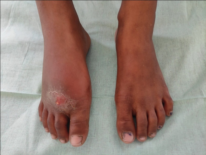
On examination, the patient was well-nourished. A diffuse swelling was noted at the anterior aspect of the forefoot and midfoot. Mild tenderness was present at the medial aspect of the junction of forefoot and midfoot; the tenderness was marked at the base and along the first metatarsal. A single discharging sinus was present over the dorsum of the right foot between the first and second metatarsals. The sinus had an opening and purulent discharge was oozing out of it; the skin around the sinus was shiny, hyperpigmented, and excoriated. Ankle dorsiflexion, plantarflexion, subtalar inversion, and eversion movements were within normal ranges. No abnormalities were detected in other joints, and the systemic examination was within normal limits with no fever, lymphadenopathy, or unremarkable respiratory, cardiovascular, or abdominal examinations.
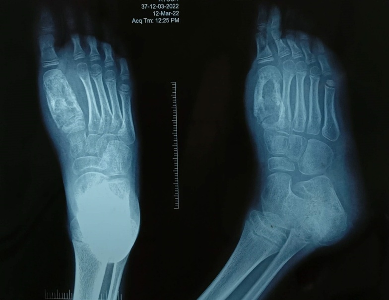
On admission, routine blood investigations like complete blood count, erythrocyte sedimentation rate, bleeding time, clotting time, liver function test, kidney function test and viral markers were sent. His erythrocyte sedimentation rate was mildly raised, but his white blood cell count and other blood tests were within the normal range. A tuberculin skin test was positive, and a chest radiograph was normal. A radiograph of the right foot revealed a centrally placed lytic lesion with an eccentrically located sclerotic lesion in the first metatarsal. The cortices of the first metatarsal were thickened, and the diaphysis was wider than in the other metatarsals. Magnetic resonance imaging study of right foot revealed an altered signal intensity of the first metatarsal, corresponding to the osteolytic lesion with intraosseous abscess, loss of cortex at places and periosteal reaction with fluid signal intensity along the first metatarsal; fluid signal intensity was also noted in the surrounding soft tissue over dorsum and plantar aspect of foot.
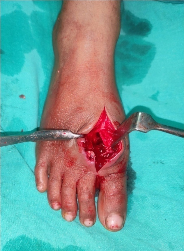
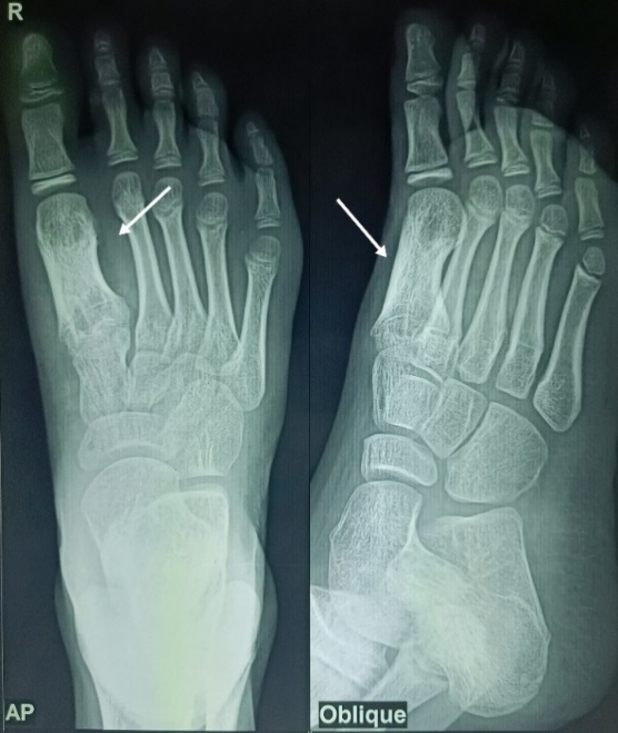
The patient was managed with curettage through a small cortical window on the superior aspect of the first metatarsal. On curettage a yellowish substance mixed with pus from dead bones, slough, and blood was obtained and was sent for culture and histopathological examination. The wound was thoroughly irrigated with normal saline. The cortical window on the superior aspect of the first metatarsal was left open for drainage and to prevent further collection. Intravenous antibiotics injection ceftriaxone were continued in the postoperative period for one week. The tuberculous bacillus was identified microscopically and microbiologically. The Ziehl–Neelsen staining showed acid-fast bacilli. Histopathological examination revealed a pyogenic granuloma with giant cells consistent with tuberculous osteomyelitis. The microbiological culture, combined with polymerase chain reaction, confirmed the presence of Mycobacterium tuberculosis. Once the diagnosis of tubercular osteomyelitis was confirmed, a long-term 18 months course of antitubercular drugs was started according to INDEX TB guidelines, which serves as the basis of the treatment. During the follow-up period, Erythrocyte sedimentation rate and CRP were reduced to normal, the pain and swelling improved promptly, and the patient was able to walk without pain. A radiograph showed remineralization of bone at 10 weeks. At the final follow-up visit, approximately 18 months after the operation, the patient remained asymptomatic without pain or deformity and did not show any local or systemic signs or symptoms suggestive of recurrence. Follow up radiograph showed bone healing through gradual disappearance of the radio-lucencies involving the first metatarsal.
Discussion
Osteoarticular tuberculosis is a relatively rare disease and accounts for only 1–3% of all tubercular infections.[3], [4] The most common locations of tuberculous osteomyelitis are the spine, femur, tibia, and fibula. [2] Approximately 10% of osteoarticular tuberculosis is known to afflict the ankle and foot, [17] making it a very uncommon condition. It typically involves the calcaneus, talus, metatarsals, navicular, and cuneiforms. [3], [4] The calcaneum is the most frequently affected bone. The plausible explanations for this include the fact that the calcaneum is the largest bone (which allows for earlier detection of lesions that are still localized to it, as opposed to the smaller bones where articular penetration into the joints is early) and there is increased susceptibility to direct trauma. [25] Isolated tubercular lesions of the metatarsal bone are rare, as reflected in the literature (Table 1). [14], [10], [11] Less than 0.5% of individuals develop metatarsal tubercular osteomyelitis. [17] With regard to metatarsal bones, tubercular infection most frequently affects the first metatarsal and least frequently the fifth metatarsal. [16]
Although up to 50% of patients do not exhibit any pulmonary manifestations, as in the present case, tuberculous osteomyelitis of the metatarsals is typically secondary to lymphohematogenous dissemination from a pulmonary lesion. [26], [27] Granulomatous foci near a joint may be the first sign of tuberculous osteomyelitis of the foot. There are four major types of tuberculosis of the ankle and foot: periarticular granuloma, central granuloma, primary haematogenous synovitis, and tenosynovitis or bursal tuberculosis. [3], [4] The periarticular granuloma, which is the most typical presentation, gradually spreads to the nearby joint if left untreated. As a result, the prognosis may substantially worsen. The second type of presentation is a central granuloma, which is quite uncommon and is more typically observed in the phalanx or metatarsal in children. [3], [4]
Metatarsal tuberculosis has a gradually progressive course. Due to the atypical appearance of disease; and ambiguous signs and symptoms, diagnosis is frequently delayed, and the disease is identified at a more advanced stage than in other forms of tuberculosis [11]. Delays frequently trigger the inexorable progression of the disease, with the adjacent joints taking the first casualty and the weight-bearing sections of the foot consequently suffering from mobility loss and deformity. [4]
The diagnosis of tuberculous osteomyelitis is challenging; it depends on a thorough history and clinical examination, radiologic findings, laboratory testing, and microbiologic and histological features. The classical presentation with regional pain, fever, anorexia, and weight loss is rarely observed, and the diagnostic conundrum gets worse if radiological presentations are unusual, especially in low incidence regions such as the foot. [28]
Clinical features consist of localized pain, edema, and muscular atrophy. Skin ulceration, discharge, difficulty in weight bearing, and joint stiffness may be observed in long-standing cases. [11] Along with the calcaneum and fifth metatarsal, the first metatarsal aids in weight bearing and is a component of the medial longitudinal arch of the foot. Pathologic involvement of the first metatarsal limits mobility during the gait cycle due to inadequate weight bearing on the afflicted bone. [29] Clinically, these people are known to have difficulty on walking on uneven ground. The index patient had difficulty on walking as well. Due to weight bearing on the afflicted bone, patients are also susceptible to pathological fractures. In the present case, the patient presented with atypical signs and symptoms at an atypical location, creating a diagnostic dilemma.
Chest X-ray screening is less beneficial for musculoskeletal tuberculosis patients because only about one-third of them have pulmonary involvement. [30] Although skin tests using a purified protein derivative are frequently positive in cases of tubercular osteomyelitis, these tests occasionally produce false-negative results.
There are no radiological features that are unique to bone TB.[2], [3], [4] Radiologic features of tuberculous osteomyelitis include osteoporosis, bone expansion with reactive sclerosis, periosteal new bone formation, increased adjacent soft tissue density, and progressive joint destruction. The characteristic appearance of cystic expansion of the short tubular bones, also known as "spina ventosa", is also a hallmark of the disease. [3], [4], [16], [31], [32]
Computed tomography scans and magnetic resonance imagings are crucial for making an early diagnosis of skeletal tuberculosis. The bony structure and geographic extent of bone deterioration, sequestration, cortical breach, and collapse are identified using computed tomography scan. [3], [4], [31], [32]
MRI is the preferred radiological test because it can detect abnormalities in and around the bone before they are visible on standard radiographs. MRI typically demonstrates an intense marrow edema with a high signal in T2-weighted images. Early MRI findings include cortical breaks, cavitation, and soft tissue collections. synovitis, joint effusions, tenosynovitis, and myositis in the surrounding tissue may be present. [3] On an MRI, the presence of an abscess aids in distinguishing between tuberculosis and a bone tumor. [4] Tuberculosis should always be considered in the differential diagnosis of a cystic lesion of the bone with hazy borders and little surrounding sclerosis.
Five radiological forms of tuberculosis of the foot are namely cystic, rheumatoid, subperiosteal, kissing, and spinaventosa by Mittal et al. These features, however, are not unique to tuberculosis and can also be present in other conditions, such as chronic pyogenic osteomyelitis and sarcoidosis. The cystic variant is relatively rare; it can be solely an intraosseous lesion, with or without the soft tissue component, and because it has not yet infiltrated the nearby joints, the prognosis after prompt initiation of therapy at this stage is favourable. Surprisingly, because other, more prevalent disease pathologies are considered first, these lesions are typically the ones that are frequently diagnosed later. [6], [17]
Because MRI findings are non-specific, a tissue diagnosis should be sought if tuberculosis is a possibility. Microbiologic and histopathological examination of the affected tissues remains the gold standard for diagnosis. FNAC is used because it supports the diagnosis and is less expensive than MRI and CT. [2] Because osteoarticular tuberculosis is paucibacillary, it is unusual to find tuberculous bacilli with Ziehl-Neelsen staining, and the isolation and culture of Mycobacterium tuberculosis remain challenging. Therefore granulomatous histological appearance and high clinical suspicion are used to make a conclusive diagnosis.[3], [4] In the present case, the tuberculosis bacillus was identified microscopically as well as genetically (using PCR).
Despite having a high degree of suspicion for tuberculous infection, bone involvement in seclusion can be confusing, and delays in diagnosis are frequent.[28] The probable reasons for this include the lack of constitutional symptoms commonly associated with osteoarticular tuberculosis elsewhere,[4], [28] the misinterpretation of radiographic characteristics with bone tumours, [3], [28] and a reasonably normal laboratory investigation picture. Despite continuous efforts, a single diagnostic test with high sensitivity and specificity for bone tuberculosis has remained elusive, and treatment is frequently started on the basis of clinical suspicion in endemic locations.[6] However, before using such a strategy, a full list of diagnostic investigations must be done, especially in cases with atypical presentations or developing in unusual locations. [3], [28]
After the development of modern multidrug chemotherapy, the prognosis for patients with tuberculosis has substantially improved. Because of the higher risk of recurrence with shorter courses of therapy, chemotherapy should last at least 12 months.[2] The fact that osteoarticular tuberculosis is a paucibacillary infection and many of the organisms are dormant and therefore resistant to chemotherapy justifies this lengthy course of treatment. [2] To avoid the collapse of articular surface, splintage and limited weight bearing may be advised.
Surgery should only be performed on patients who do not respond to conservative management or as a supplement to medical therapy.[3], [4] In cases of metatarsal TB, surgical options include open biopsy in dubious cases and curettage of juxta-articular cavities with or without debridement. A lesion that is near the articular surface should be taken into consideration for debridement because performing so may prevent further progression and joint invasion and help avoid worsening of the prognosis.[3], [4], [25] This might also apply to some osteolytic foot lesions that are superficial and easy-to-operate and leave little to no residual post-op disability.[25] In the present case, the patient responded well to medical therapy after curettage of the lesion.
It has been clearly established that the radiographic image does change after treatment.[28] Cavities often take a very long time to disappear, and minor residual cavities may still be evident years after therapy but are of little clinical importance.[28] Unless other nearby bone or joint is affected, the prognosis is typically favourable.[17]
Conclusion
Tuberculosis is a common problem in India. It can affect any part of the body, but rarely involves the foot. Tuberculosis of the metatarsal is a difficult diagnosis due to its rarity and atypical presentation. In areas with a high prevalence of tuberculosis, there may be no history of tubercular contact, or any ‘typical’ symptoms or signs.
A high index of clinical suspicion is required for early diagnosis which can prevent inevitable spread of the disease to adjacent joints. It is important to recognize the less common presentations of this condition to enable early diagnosis and successful treatment. Prompt non-invasive investigations such as CT and MRI should be performed to detect early lesions. Because of the multiple possible clinical diagnoses, radiographic and histopathological assessments are very critical for making an accurate diagnosis and initiating treatment of tuberculous osteomyelitis.
Prolonged antitubercular therapy (12-18 months) along with protected weight bearing provides good clinical results even in delayed cases. Debridement and curettage of the affected bone, rather than en bloc amputation, is usually recommended, particularly when soft tissue granuloma and ulceration are present. Resection of the entire diseased metatarsal is rarely essential, as evidenced by satisfactory long-term recovery of our patient; however a long-term surveillance is required.
Research Ethics and Patient Consent
Written consent for publication of patient details were obtained from parents.
Source of Funding
None.
Conflict of Interests
All the authors declare that there is no conflict of interest.
References
- Nayak B, Dash R, Mohapatra K, Panda G. Ankle and foot tuberculosis: a diagnostic dilemma. J Fam Med Primary Care. 2014;3(2):129-31. [Google Scholar]
- Tuli S. . Tuberculosis of the skeletal system. 4th edn. . 2010. [Google Scholar]
- Dhillon M, Sharma S, Gill S, Nagi O. Tuberculosis of bones and joints of the foot: an analysis of 22 cases. Foot Ankle. 1993;14(9):505-13. [Google Scholar]
- Dhillon M, Nagi O. Tuberculosis of the foot and ankle. Clin Orthop Relat Res. 2002;398:107-13. [Google Scholar] [Crossref]
- Watts H, Lifeso R. Current concepts review. Tuberculosis of bones and joints. J Bone Joint Surg Am. 1996;78(2):288-98. [Google Scholar]
- Mittal R, Gupta V, Rastogi S. Tuberculosis of the foot. J Bone Joint Surg. 1999;81(6):997-1000. [Google Scholar]
- Reading A, Stother I. The painless fracture: could it be TB?. J R Coll Surg Edinb. 1998;43(6):410-1. [Google Scholar]
- Negusse W. Bone and joint tuberculosis in childhood in a children’s hospital Addis Abeba. Ethiop Med J. 1993;31(1):51-61. [Google Scholar]
- Lonner J, Sheskier S. Tuberculosis of the foot as the initial manifestation of acquired immune deficiency syndrome: a report of two cases. Foot Ankle Int. 1995;16(3):167-71. [Google Scholar]
- Vohra I, Kang H, Dogra S, Saggar R, Sharma R. Tuberculous osteomyelitis. J Bone Joint Surg Br. 1997;79(4):562-6. [Google Scholar]
- Muratori F, Pezzillo F, Nizegorodcew T, Fantoni M, Visconti E, Maccauro G. Tubercular osteomyelitis of the second metatarsal: a case report. J Foot Ankle Surg. 2011;50(5):577-9. [Google Scholar]
- Vijay V, Sud A, Mehtani A. Multifocal bilateral metatarsal tuberculosis: a rare presentation. J Foot Ankle Surg. 2015;54(1):112-5. [Google Scholar]
- Jain V, Kumar D, Arya R, Sinha S, Naik A. Tubercular osteomyelitis of the first metatarsal bone as a cause of forefoot pain. Foot (Edinb). 2016;27:19-21. [Google Scholar] [Crossref]
- Sarwal S, Singh M, Gautam R, Gill S, Hunjan J, Arora M. Tubercular osteomyelitis of 1st metatarsal bone right foot. Int J Scientific Res. 2016;5(10):64-6. [Google Scholar]
- Apoorva H, Jain A, Kumar S. Tuberculosis of fifth metatarsal bone- a rare case report. Med Sci. 2019;8(2):457-9. [Google Scholar]
- Pes M, Amorese V, Baioni A, Donadu M, Molicotti P, Milia F. A rare presentation of tubercular osteomyelitis of the foot. J Infect Dev Ctries. 2022;16(10):1655-9. [Google Scholar]
- Yasser B, Khalid A. Isolated first metatarsal Tuberculous osteomyelitis in a pediatric patient: A Case Report. JMCRR. 2023;6(3):1250-4. [Google Scholar]
- Yadav S, Rawal G, Jeyaraman M. Primary tuberculosis of the cuboid and fifth metatarsal without pulmonary involvement: A rare case report. Cureus. 2023;15(8). [Google Scholar] [Crossref]
- Ganda V, Kadu V, Gadghate N, Saindane K. Unusual site of tubercular osteomyelitis of fifth metatarsal. Int J Health Sci Res. 2015;5(2):479-82. [Google Scholar]
- Madi S, Naik M, Vijayan S. An isolated case of first metatarsal tuberculosis. Oxf Med Case Rep. 2015;3:241-3. [Google Scholar] [Crossref]
- Prakash J, Agnihotri A, Jaiswal Y, Mehtani A. A very rare cause of chronic foot pain in a child: metatarsal tubercular osteomyelitis. BMJ Case Rep. 2014. [Google Scholar] [Crossref]
- Yuen M, Tung W. An uncommon cause of foot ulcer: tuberculosis osteomyelitis. Emerg Med J. 2001;18(2):140-1. [Google Scholar]
- Jana P, Das I, Sanyal D, Basuthakur S, Mandal K. Tuberculosis of 4th and 5th metatarsals bone of an adult patient. Int Med J. 2006;4(13):297-8. [Google Scholar]
- Flint J, Saravana S. Tuberculous osteomyelitis of the midfoot: a case report. Cases J. 2009;2. [Google Scholar] [Crossref]
- Dhillon M, Aggarwal S, Prabhakar S, Bachhal V. Tuberculosis of the foot: An osteolytic variety. Indian J Orthop. 2012;46(2):206-11. [Google Scholar]
- Vallejo J, Ong L, Starke J. Tuberculous osteomyelitis of the long bones in children. Pediatr Infect Dis J. 1995;14(6):542-6. [Google Scholar]
- Huang C. Extra-articular tuberculous osteomyelitis. A report of 11 cases. Int Orthop. 1996;20(3):169-71. [Google Scholar]
- Dhillon M, Tuli S. Osteoarticular tuberculosis of the foot and ankle. Foot Ankle Int. 2001;22(8):679-86. [Google Scholar]
- Dhillon MS, Aggarwal S, Prabhakar S, Bachhal V. Tuberculosis of the foot: An osteolytic variety. Indian J Orthop. 2012;46:206-217. [Google Scholar]
- Daniel T, Debanne S. The serodiagnosis of tuberculosis and other mycobacterial diseases by enzyme-linked immunosorbent assay. Am Rev Resp Dis. 1987;135(5):1137-51. [Google Scholar]
- Choi W, Han S, Joo J, Kim B, Lee J. Diagnostic dilemma of tuberculosis in the foot and ankle. Foot Ankle Int. 2008;29(7):711-5. [Google Scholar]
- Korim M, Patel R, Allen P, Mangwani J. Foot and ankle tuberculosis: case series and literature review. Foot (Edinb). 2014;24(4):176-9. [Google Scholar]